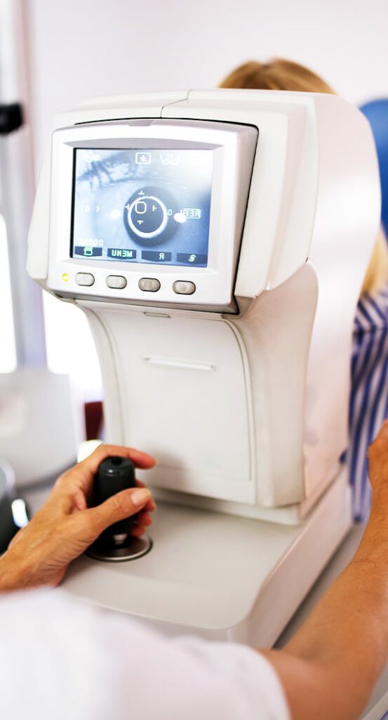
Diagnostic Eye Care Technology

We use diagnostic technology at Toronto Medical Eye Associates to help provide our patients with an excellent and effective eye care experience.
Our diagnostic technology can help us get a clearer picture of our patient’s eyes and determine irregularities and the development of eye diseases.
Contact us to book your comprehensive eye exam and get your expert diagnosis done today.
Comprehensive Vision Care
Our goal at Toronto Medical Eye Associates is to provide comprehensive eye exams using effective diagnostic technology to provide every angle possible for eye care.
Your eye health and visual needs are a priority, and diagnosing eye diseases early can be essential to prevent further damage and prepare an effective treatment plan.

Explore Our Diagnostic Services
We offer advanced diagnostic services to assist with eye exams, eye disease management, and other vision care issues.
Retinal Imaging
Annual eye exams are vital to maintaining your vision and overall health. We offer Optomap retinal imaging as an essential part of our eye exams.
The optomap produces a unique image and provides our doctors with a high-resolution 200° image to determine the health of your retina, which is much wider than a traditional 45° image.
Many eye problems can develop without you knowing, and you may not even notice a change in your sight. Fortunately, diseases or damage such as macular degeneration, glaucoma, retinal tears or detachments, and other health problems such as diabetes and high blood pressure can be seen with a thorough retina exam.
The inclusion of Optomap retinal imaging as part of a comprehensive eye exam provides:
- A scan to show a healthy eye or detect diseases.
- A view of the retina, giving your doctor a more comprehensive view than they can get by other means.
- The opportunity for you to view and discuss the optomap image of your eye with your doctor at the time of your exam.
- A permanent record for your file, which allows our doctors to view your images each year to look for changes.
Optomap retinal imaging is fast, easy, and comfortable for our patients. The entire imaging process consists of you looking into the device one eye at a time. The optomap images are then displayed on a computer screen so we can review them with you.
Optical Coherence Tomography with Angiography Capability
At Toronto Medical Eye Associates, we offer AngioPlex OCT Angiography from ZEISS to provide non-invasive imaging of retinal microvasculature. This diagnostic technology helps take glaucoma and retinal disease management and treatment planning to the next level.
OCT Angiography reveals early indicators and offers advanced care for many ocular diseases, including diabetic retinopathy, age-related macular degeneration (AMD), glaucoma, and more.
Heidelberg Retinal Tomography (HRT) Technology
Heidelberg retinal tomography is a diagnostic procedure used for precise observation and documentation of the optic nerve head, which is essential for diagnosing and managing glaucoma.
The HRT uses a special laser to take 3D photographs of the optic nerve and surrounding retina. This laser, which will not harm the eye, is focused on the surface of the optic nerve and captures the image.
The HRT takes images of deeper layers until the desired depth has been reached. Finally, the instrument takes these pictures of the layers and combines them to form a 3D image of the entire optic nerve.
Typical optic nerve damage in glaucoma is known as “cupping.” As the cells that make up the nerve are damaged due to increased pressure inside the eye, they die and disappear. When a large number of these cells are gone, they leave behind a small “cup” in the nerve, so our doctors look for this when they examine the optic nerve to see how deep and wide it is.
The HRT image can be used to determine the area of the optic disc, the volume of the cup, and the area of the rim around the cup. Over several visits, scans are layered, and changes are measured.
Humphrey’s Visual Field
A Humphrey’s visual field test is used to help your doctor detect and treat glaucoma and diagnose possible neurological conditions such as pituitary tumours, optic neuritis, stroke, and more.
A visual field maps out your peripheral vision using a center fixation light and blinking lights. The patient is instructed to always focus on the center fixation light and press the button when the blinking test light is seen with your side vision.
If you move your eyes to follow or look for the blinking lights, it decreases the reliability of the test and the ability of your doctor to detect and/or monitor your disease. Visual field testing is crucial in detecting eye diseases that may have otherwise gone undetected. Often, subtle changes in our peripheral vision cannot be detected except through this highly sensitive and validated test.
Visual field testing can detect the most subtle visual field changes that a patient would very likely never notice. In the instance of glaucoma progression, your doctor can initiate treatment in an attempt to prevent further peripheral vision loss.
When glaucoma advances, it continues to impact peripheral vision until the disease is in its end stages, where it may lead to blindness. Early detection is crucial, and we use advanced diagnostic tools in an effort to preserve your vision.
Meibography & Dry Eye Analysis
Meibography is a specialized imaging technology designed to visualize the meibomian glands’ morphology and can assist with dry eye analysis.
These tools offer non-invasive tear break-up time (NIBUT), meibomian gland imaging with the area of:
- Loss analysis
- Tear meniscus height analysis
- Blink analysis
- Real fluorescein imaging and video acquisition
- View review of anterior corneal aberrations between blinks
- Tear lab
- Inflammadry
Corneal Topography & Axial Length
Corneal topography analyzes the anterior cornea, including topographic maps, 3D maps, comparison maps, height maps, Zernike analysis, and keratoconus screening.
Corneal topography can help us examine characteristics of the cornea such as shape, curvature, power, and thickness. It is also an essential tool for contact lens fitting.
The axial length of the eye refers to the length of the eye from front to back. Measuring the axial length of the eye can indicate the speed at which a patient’s myopia is progressing and can help us determine the cause of the eye’s elongation.
Axial length measurements are key in developing and monitoring myopia management treatments for young children.
We Care About the Details
At Toronto Medical Eye Associates, our team values the details, and we use diagnostic technology to get a clear picture of your eyes and your unique vision needs.
Contact us today to book your comprehensive eye exam and get a detailed picture of your vision situation.
Our Services


Say Goodbye to Dry Eye
Do you suffer from dry eye symptoms?
Dry eye is a common condition that can cause your eyes to feel uncomfortable and irritated. Don’t let dry eye stop you from living your life. Dr. Taji is a trusted voice on dry eye disease and has hosted many lectures and presentations on diagnosis and treatment.
You can feel confident in your dry eye treatment with our team. Contact us today to book your dry eye evaluation and get relief sooner.
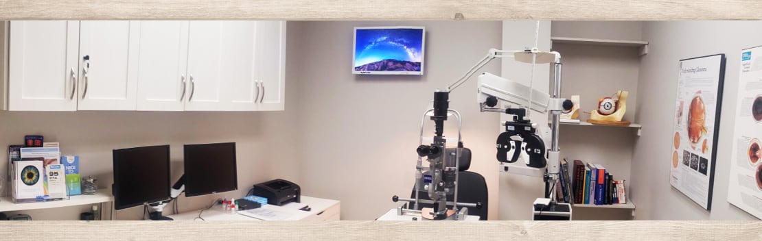
Our Location
You’ll find our clinic around the corner from North York General Hospital, just up from the Don River and Betty Sutherland hiking trail.
Please don’t hesitate to contact us if you have any trouble finding us!
Our Address
- 1333 Sheppard Avenue East, Suite 343
- North York, Ontario M2J 1V1
Contact Information
- 416-494-2020
- 416-494-0010
- info@torontomedeye.ca
Our Hours
- Monday – Thursday: 8:30 AM – 5:00 PM
- Friday: 8:30 AM – 12:00 PM
- Saturday & Sunday: Closed

See Our Google Reviews
Eye Exams
Specialty Eye Exams
Optical
Toronto Medical Eye Associates
- 1333 Sheppard Avenue East, Suite 343
- North York, Ontario M2J 1V1
- 416-494-2020
- 416-494-0010
- info@torontomedeye.ca